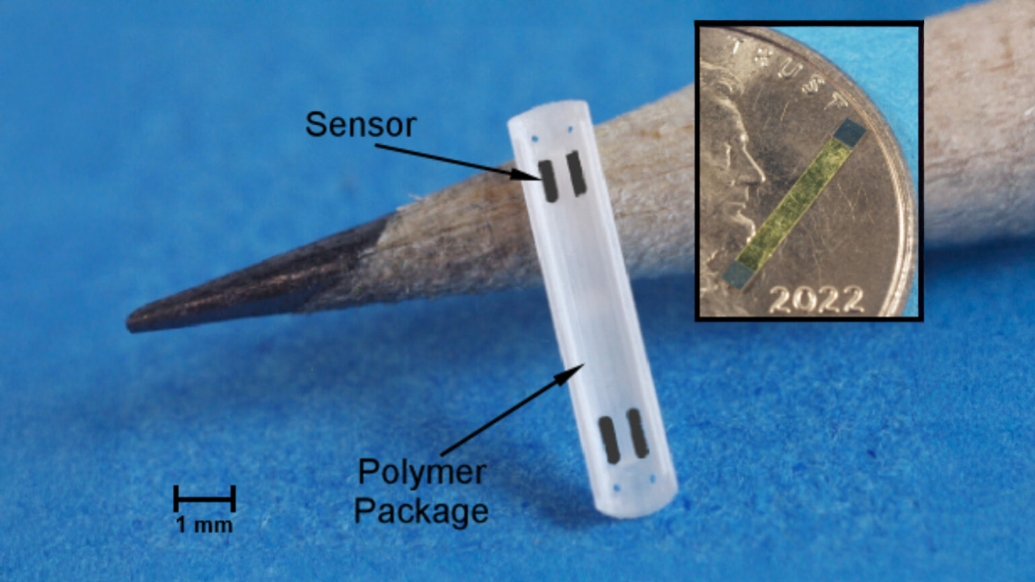
New battery-less and wireless sensor tested in pigs.

Stents to treat various blockages in the human body can themselves become blocked, but a new sensor developed at the University of Michigan for stents that are used in the bile duct may one day help doctors detect and treat stent blockages early, helping keep patients healthier.
Bile duct blockages can cause jaundice, liver damage, and potentially life-threatening infections.
Conditions that cause the bile ducts to narrow and close, including pancreatic and liver cancers, may be treated by inserting stents to prop the ducts open.
However, bacterial sludge or gallstones can block bile duct stents, requiring urgent treatment with antibiotics and stent replacement.
Currently, medical providers monitor biliary stent blockages through blood tests, meaning the problem must be significant enough for the body to notice.
A sensor within the stent could enable doctors to recognize that bacterial sludge is accumulating and intervene before the patient begins to fall ill.
“This novel stent sensor provides the opportunity to detect impending biliary stent obstructions without waiting for clinical symptoms, blood tests or imaging tests, all of which delay intervention,” said Richard Kwon, a clinical professor of internal medicine and gastroenterology at the U-M Medical School and co-author of the study in Nature Microsystems & Nanoengineering.
The sensor is 8 millimeters long, a little less than half the diameter of a penny, and only 1 millimeter wide, encapsulated in a protective, 3D-printed plastic structure that bonds to plastic stents.
During a checkup, the patient would don a belt-like detector around the waist that emits an alternating magnetic field, changing sign at different frequencies to induce the maximum, or resonant, vibration in the sensor.
As the sensor vibrates, any masses weighing down the sensor show up in a lowered resonant frequency.
“Successfully receiving signals from a live animal marks a major advancement in low profile, batteryless magnetoelastic sensor technology, paving the way for new and expanded applications,” said Ramprasad Nambisan, U-M doctoral graduate of electrical and computer engineering and lead author of the study.
One of the big challenges was detecting that resonant frequency, which shows up as an answering magnetic field coming from the sensor, even through nearly 7 inches of fluid-rich abdominal tissue.
With careful hardware design and digital signal processing, the team registered a signal-to-noise ratio of a million to one during testing.
“The high value measured for the signal-to-noise ratio at 17 cm interrogation distance indicates that in the future the readout distance can be greatly increased as necessary for humans, accommodating differences in anatomy,” said Yogesh Gianchandani, U-M professor of electrical and computer engineering and senior author of the study.
As next steps, the researchers plan to develop a version that works with metal stents.
In the longer term, they will further miniaturize the sensor—allowing several sensors to be distributed along the stent, each with a different resonant frequency.
This would enable localized detection of sludge accumulation.
The team also intends to develop lower-cost electronics for the belt-like detector, paving the way for human clinical trials.
As the technology continues to advance, magnetoelastic sensors have the potential to be used in other places in the body, including peripheral vascular stents, long-term coronary stents, and ureteral stents.
This research includes co-authors at Michigan Medicine and was funded in part by the National Institutes of Health (R01DK102663).
The device was built in part using the facilities in the Lurie Nanofabrication Facility and studied at the Michigan Center for Materials Characterization.
The team has applied for patent protection with the assistance of U-M Innovation Partnerships and is seeking partners to bring the technology to market.
Study: “A microsystem for in vivo wireless monitoring of plastic biliary stents using magnetoelastic sensors,” Nature Microsystems & Nanoengineering. DOI: 10.1038/s41378-024-00772-8.
This story was previously published on the Michigan News website on October 31, 2024, and by the Michigan Medicine Health Lab Blog on November 22, 2024.